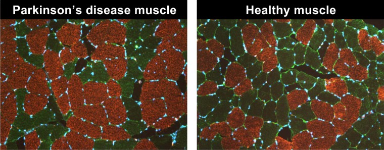
Fig. 1.
Representative images of type I myofiber grouping in skeletal muscle biopsy specimens collected from the vastus lateralis of individuals with Parkinson’s disease in comparison to healthy skeletal muscle. Our laboratory’s methodology for quantification of type I myofiber grouping is presented briefly in methods and in greater detail elsewhere (37), and we have previously investigated the potential implications of type I grouping in Parkinson’s disease (36).

