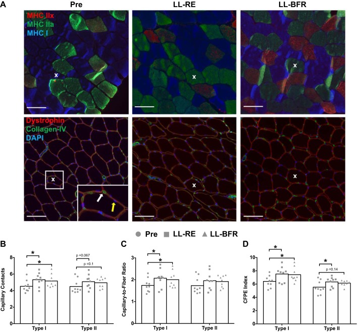
Fig. 4.
Skeletal muscle microvascular properties before (Pre) and after 6 wk of low-load resistance exercise (LL-RE) or low-load blood flow restriction resistance exercise (LL-BFR) training. A: representative images for immunohistochemical staining of serial cross sections. Top: type I [myosin heavy chain (MHC) I, blue] and type II (MHC IIa, green; MHC IIx, red) muscle fibers. Bottom: muscle fiber boarder (dystrophin; red), capillary (collagen IV, green), and nuclei (DAPI, blue). White and yellow arrows represent a capillary and a myonucleus, respectively. X depicts the same fiber within each condition for both sets of images. Scale bars = 100 μm. B–D: capillary contacts, capillary-to-fiber ratio, and capillary-to-fiber perimeter exchange (CFPE) index. *P < 0.05 vs. Pre. Symbols represent individual values, and bars represent group means (n = 9/group).

