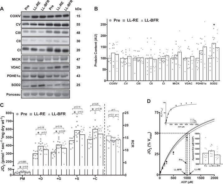
Fig. 5.
Mitochondria-located protein expression and respiratory capacity in permeabilized muscle fibers before (Pre) and after 6 wk of low-load resistance exercise (LL-RE) or low-load blood flow restriction resistance exercise (LL-BFR) training. A: representative Western blot images for each protein for 2 participants. COXIV, cytochrome c oxidase complex IV; CI–CIII and CV, complexes I–III and V; MiCK, mitochondrial creatine kinase; VDAC, voltage-dependent anion-selective channel protein; PDHE1α, pyruvate dehydrogenase E1 α-subunit. B: quantification of Western blots for proteins located within mitochondria. AU, arbitrary units. C: high-resolution respirometry illustrating maximal complex I/II-linked respiration. D: Michaelis-Menten kinetic curves and apparent Km for ADP. Jo2, O2 consumption; PM, pyruvate + malate; +D, PM + ADP; +G, PMD + glutamate; +S, PMDG + succinate; +C, PMDGS + cytochrome c; RCR, respiratory control ratio. *P < 0.01 vs. Pre. Symbols represent individual values, and bars represent group means [n = 7–10/group (Western blot data) and n = 10/group (respirometry data)].

