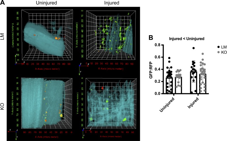
Fig. 7.
Autophagy flux is reduced in mice after traumatic freeze injury. A: representative two-photon microscopy images from tibialis anterior (TA) muscles of littermate (LM) and Unc-51 like autophagy activity kinase (ULK) muscle-specific knockout (MKO) mice transfected with green fluorescent protein (GFP)-microtubule-associated protein light chain B1 (LC3)-red fluorescent protein (RFP)-LC3ΔG plasmid. B: quantitative analysis of the GFP:RFP ratio which represents autophagosome turnover. Autophagy flux was greater in uninjured muscle as opposed to injured muscle. Data are presented as individual values ± SD. Differences were analyzed by two-way RM ANOVA with the repeated measures being the injured vs. uninjured contralateral control limb and the other factor being genotype. Main effects are reported.

