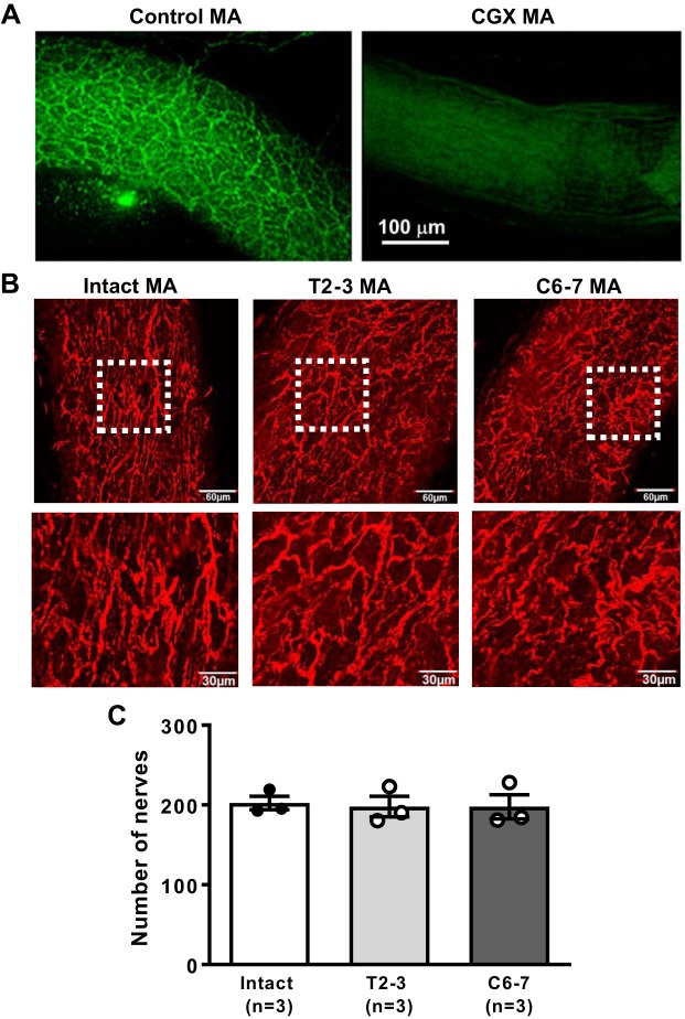
Fig. 7.
Comparison of tyrosine hydroxylase (TH) immunoreactive nerves supplying mesenteric arteries (MA) of intact, T2–3, and C6–7 rats. A: immunohistochemical labeling of TH-positive sympathetic nerves supplying a MA in the small intestine. Left: TH labeling of a MA from a control rat shows a dense nerve plexus. Right: TH-positive nerves are absent from a MA from a rat 7 days after celiac ganglionectomy (CGX). Scale bar applies to both images. B: representative images showing the distribution of sympathetic nerve fibers on the adventitial surface of MA. C: nerve fiber grid crossing counts show that the nerve density was similar in MA from intact, T2–3, and C6–7 rats.

