Fig. 3.
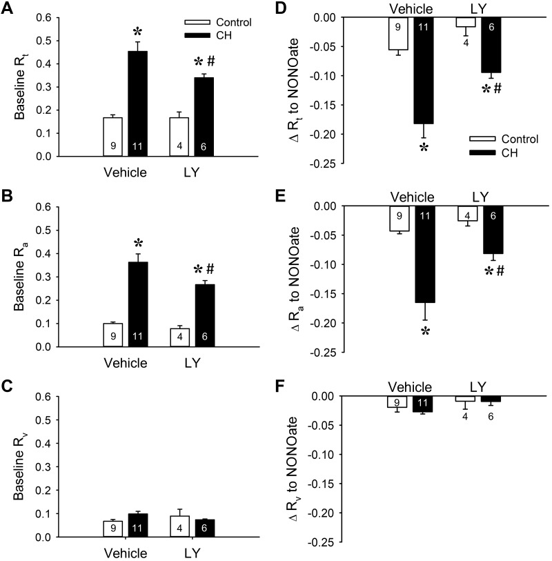
Chronic hypoxia (CH) increases baseline pulmonary vascular resistance and basal tone through PKCβ signaling. A–C: total (Rt; A), arterial (Ra; B), and venous (Rv: C) baseline vascular resistance (mmHg·mL−1·kg·min) in lungs (in situ) from control and CH neonatal rats in the presence or absence of the PKCβ inhibitor LY-333,531 (LY, 10 nM). D–F: the contribution of basal tone to total (D), arterial (E), and venous (F) resistance is expressed as the change in resistance (ΔR) to 1,3-propanediamine, N-{4-[1-(3-aminopropyl)-2-hydroxy-2-nitro-sohydrazino]butyl} (spermine NONOate, 100 μM) in lungs from control and CH neonates. Experiments were conducted in the presence of Nω-nitro-l-arginine (300 μM). Values are means ± SE; n = 4–11 rats/group (indicated in bars). *P < 0.05 vs. control, #P < 0.05 vs. vehicle, analyzed by 2-way ANOVA followed by Student-Newman-Keuls post hoc comparison.

