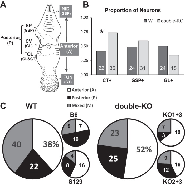
Fig. 7.

Receptive fields of recorded neurons. A: schematic of the mouse oral cavity depicting locations of the different taste receptor subpopulations and their innervation. Lingual taste buds include the fungiform papilla on the anterior tongue (FUN) innervated by the chorda tympani (CT) branch of the 7th nerve, and foliate (FOL), and circumvallate (CV) papillae on the posterior tongue, mostly innervated by the glossopharyngeal nerve (GL; the CT also innervates a few foliate taste buds). Taste buds occur in 2 regions of the palate, anteriorly in the nasoincisor ducts (NID) and posteriorly on the soft palate (SP); both palatal fields are innervated by the greater superficial petrosal (GSP) branch of the 7th nerve. B and C: proportions and numbers of neurons with receptive fields classified by innervation or oral cavity location (receptive fields were only determined for a subset of the recorded neurons). There was a higher proportion of double-KO neurons with CT inputs, as shown by *. B: the proportions (y-axis) and numbers (shown in bars) of neurons that could be classified as receiving or not receiving inputs from a given nerve. Note that proportions do not add up to “1.” C: pie charts showing the distribution of neurons with receptive fields in the anterior mouth (A) (FUN and/or NID), posterior (P) (FOL, SP, and/or CV), and mixed (M) (both A and P). Large pie charts represent pooled data for wild-type (WT) and double-knockouts (KOs) with percentages indicated. The smaller graphs break down the distribution for the individual strains, with numbers indicating the sample sizes.
