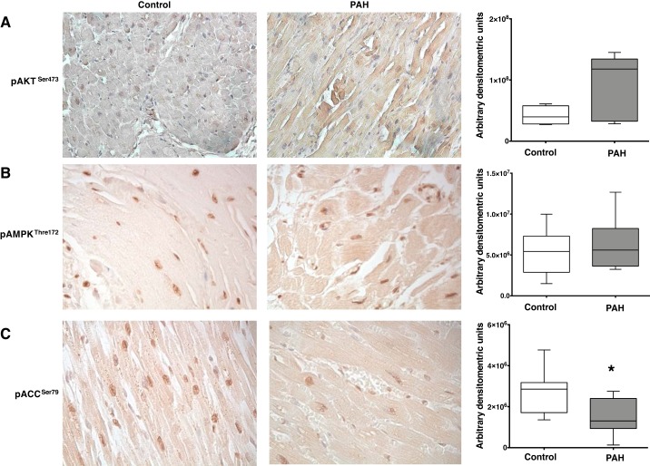
Fig. 3.
Immunohistochemical analysis of phosphorylated (p)-AktSer473, p-AMPKThr172, and p-acetyl-CoA carboxylase (ACC)Ser79 in human right ventricular (RV) tissue from control and pulmonary arterial hypertension (PAH) patients. A: pAktSer473 staining the RV tissue is in brown and nuclei were counterstained with hematoxylin. Magnification ×400. Bar graphs represent semiquantitative analysis of brown staining using Fiji software [control (n = 4), PAH (n = 6)]. B: pAMPKThr172 staining the RV tissue is in brown and nuclei were counterstained with hematoxylin. Magnification ×400. Bar graphs represent semiquantitative analysis of brown staining using Fiji software [control (n = 7), PAH (n = 8)]. C: pACCSer79 staining the RV tissue is in brown and nuclei were counterstained with hematoxylin. Magnification ×400. Bar graphs represent semiquantitative analysis of brown staining using Fiji software [control (n = 7), PAH (n = 8)]. *P < 0.05. Statistical test used was t test.

