Fig. 4.
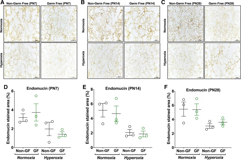
No difference in microvascular density is noticed between germ-free (GF) and non–germ-free (NGF) animals. A–C: representative photomicrographs of endomucin-stained sections of lungs from Swiss Webster (NGF and GF) mouse pups [postnatal age 7 (PN7; A), postnatal age 14 (PN14; B), and postnatal age 28 (PN28; C)] in normoxia (21% FiO2) or hyperoxia (85% FiO2) exposure from PN3–PN14 (magnification, ×200). D–F: hyperoxia exposure resulted in sparse microvasculature in both NGF and GF mice at all postnatal ages compared with their respective normoxia group on analysis using endomucin stained area (%). No difference was seen between NGF and GF groups in microvascular density in either normoxia or hyperoxia conditions. Data are means ± SE; n = 3 animals per group.

