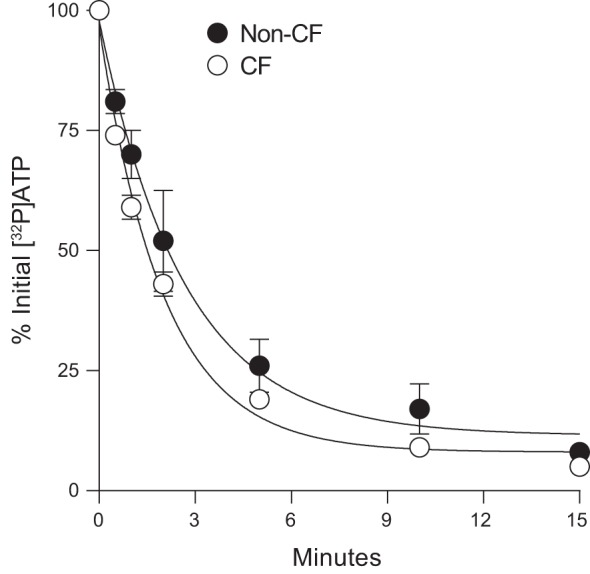
Fig. 1.

Decay of [γ32P]ATP on human bronchial epithelial cells (HBEC). Incubations were initiated by the addition of 0.1 μCi [γ32P]ATP to 250 μL HBSS bathing the luminal surface of non-cystic fibrosis (CF) and CF HBEC cultures. Aliquots were removed at the time indicated and the resulting 32P-labeled species were quantified by HPLC, as described in methods. The data represent the mean ± SD from three experiments. Exponential decay lines were generated, and first-order rate constant values were calculated (non-CF, k = 0.37 min−1; CF, k = 0.49 min−1) using Sigma Plot 10 regression fitting.
