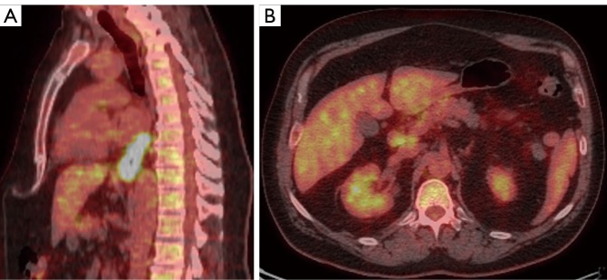
Figure 1.
PET/CT sagittal (A) and axial (B) of esophageal carcinoma. (A) Initial PET/CT with long-segment distal esophageal thickening (max SUV 18.3); (B) FDG-avid regional lymphadenopathy (partially-displayed), largest portocaval (max SUV 4.3). Note normal background liver activity. PET/CT, positron emission tomography/computed tomography; SUV, standard uptake value.

