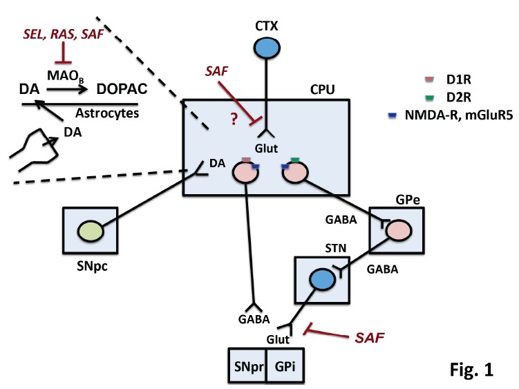
Fig. (1).

Mechanisms involved in the antiparkinsonian effect of selegiline (SEL), rasagiline (RAS), and safinamide (SAF). The Figure is a schematic drawing of the basal ganglia motor circuit, with dopaminergic neurons of the substantia nigra pars compacta (SNpc) projecting to the caudate nucleus and putamen (CPU). Striatal projection neurons of the direct and indirect pathway are stimulated and inhibited by D1 and D2 dopamine receptors (D1R, D2R), respectively. Both neurons express NMDA receptors (NMDA-R) and mGlu5 metabotroic glutamate receptors (mGluR5), which are activated by the glutamate released from cortico-striatal fibers (CTX = cerebral cortex). The external globus pallidus (GPe) and the subthatamic nucleus (STN) are the two stations of the indirect pathway. STN glutamatergic neurons send excitatory projections to the internal globus pallidus (GPi) and the substantia nigra pars reticulate (SNpr). Neurons of the direct patway send GABAergic projection to the GPi and SNpr. Dopamine (DA) released from nigro-striatal terminals is taken up by astrocytes, where it is transformed into DOPAC by MAOB. Inhibition of MAOB by SEL, RAS, and SAF enhances DA levels in the striatum. SAF can also inhibit glutamate release in the GP and SNpr. Inhibition of glutamate release in the striatum by SAF was not demonstrated by in vivo microdialysis experiments [33]. (The color version of the figure is available in the electronic copy of the article).
