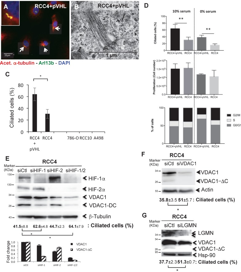
Figure 2.
The presence of VDAC1-DC in ccRCC cells decreases or abolishes ciliation. A, Triple immunofluorescence labeling and merged images with acetylated α-tubulin (Acet. α-tubulin in red), Arl13b (in green) and DAPI (in blue). B, Electron microscopy of RCC4+pVHL cells. C, Quantitative analysis of the ciliation percentage was assessed by confocal fluorescence microscopy (n=100-300 cells). D, Both cell lines were seeded at the same density and incubated in Nx for 48h with or without serum. Percentage of ciliated cells, proliferation and FACS analysis were measured. The mean ± SEM is representative of three independent experiments carried out in duplicate. E, F and G, RCC4 cells were transfected with control siRNA (siCtl), (E) siHIF-1α, siHIF-2α and siHIF-1/2α, (F) siVDAC1 and (G) siLGMN. Cell lysates were analyzed by immunoblotting for HIF-1α, HIF-2α, VDAC1, LGMN and β- tubulin/Actin or HSP90 were used as a loading control. Quantitative analysis of the ciliation percentage was assessed by confocal fluorescence microscopy (n=100-300 cells). A * p<0.05 shows significant differences. Quantification of VDAC1 and VDAC1-ΔC protein levels (E). Experiments have been proceeded without serum.

