Figure 6.
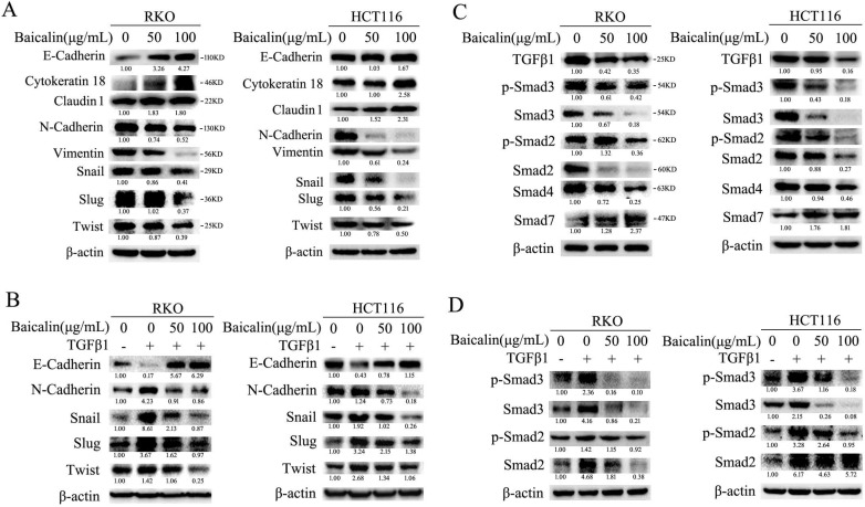
Baicalin inhibited epithelial to mesenchymal transition (EMT) by suppressing TGF-β/Smad pathway in CRC cells. (A) RKO and HCT116 cells were treated with different concentrations of baicalin for 48 h, and then cells were harvested and the protein levels of E-cadherin, Cytokeratin 18, Claudin 1, N-Cadherin, Vimentin, Snail, Slug, Twist and β-actin were determined by western blotting. (B) After treating with different concentrations of baicalin for 48 h, RKO and HCT116 cells were treated or untreated with TGF-β1 (10 ng/mL) for another 12 h; and then cells were harvested. The protein levels, including E-cadherin, N-cadherin, Snail, Slug and Twist, were detected by western blot analysis. (C) RKO and HCT116 cells were incubated with increasing doses of baicalin and total cell lysates were analyzed by immunoblotting with anti-phospho Smad2/3, anti-Smad2/3/4/7 antibody. (D) After treating with different concentrations of baicalin for 48 h, RKO and HCT116 cells were treated or untreated with TGF-β1 (10ng/mL) for another 12 h; and then cells were harvested. The protein levels of p-Smad2/3 and Smad2/3 were analyzed by immunoblotting. The relative protein levels in western-blot assay were quantified and marked under the protein bands.

