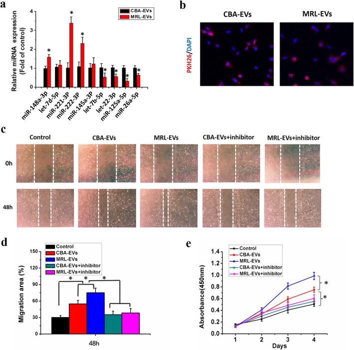
Fig. 8.
MRL-EVs promote the chondrogenesis via transferring miR-221-3p. a Quantitative real-time PCR validation of miRNAs in extracellular vesicles secreted by CPCs derived from CBA and MRL mice. *p < 0.05 represents significant differences between CBA-EVs and MRL-EVs. n = 3 per group. b Representative immunofluorescence photomicrograph of PKH26 (red)-labeled extracellular vesicles absorbed by chondrocytes, the nuclei of which were stained by DAPI (blue). c Light microscopy images of scratch wound assays. The migration of chondrocytes in different treatment groups was tested by the scratch wound assay. d Quantitative analysis of migration rates at 48 h. Both CBA-EVs and MRL-EVs promoted chondrocyte migration and that MRL-EVs were more effective than CBA-EVs, but this effect was reduced by miR-221-3p inhibitor. *p < 0.05. n = 3 per group. e Effects of CBA-EVs and MRL-EVs on proliferation of chondrocytes. CCK-8 assay showed that CBA-EVs and MRL-EVs promoted chondrocyte proliferation and that MRL-EVs were more effective than CBA-EVs, but the miR-221-3p inhibitor markedly decreased their upregulation induced by CBA/MRL-EVs. *p < 0.05. n = 3 per group

