Fig. 1.
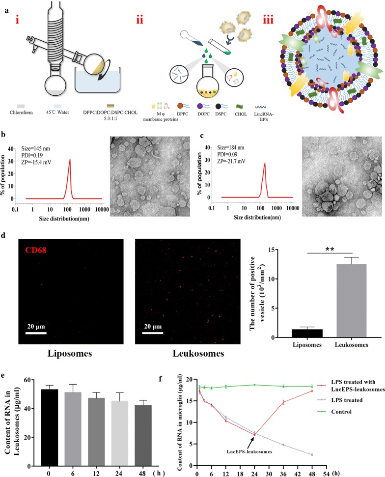
LncEPS-leukosome synthesis, formulation, and physicochemical features. a Liposome film was prepared by rotary vacuum evaporation. Extraction of membrane proteins from Mφ. Membrane proteins and lincRNA-EPS were loaded into the liposome. Pre-designed structure of LncEPS-leukosome. b, c The Dynamic Light Scattering (DLS) analysis show the size, zeta potential (ZP) and polydispersity index (PDI) of liposomes (b) and leukosomes (c). Cryo-TEM analysis show morphological of two formulations. d Immunofluorescence assay confirmed that membrane proteins were bound to the membrane of leukosomes. Liposomes and leukosomes were stained with an anti-CD86 antibody (red). e RT-PCR demonstrating the envelopment and leakage rate of lincEPS-leukosomes construed by 100 μg/ml lincRNA-EPS. f Changes in expression of microglial lncRNA-EPS after activation or treatment with lncEPS-leukosome were demonstrated by RT-PCR. Scale bar, 20 μm. (n = 8 per group). Values represent the mean ± SD, * and #P < 0.05, ** and ##P < 0.01

