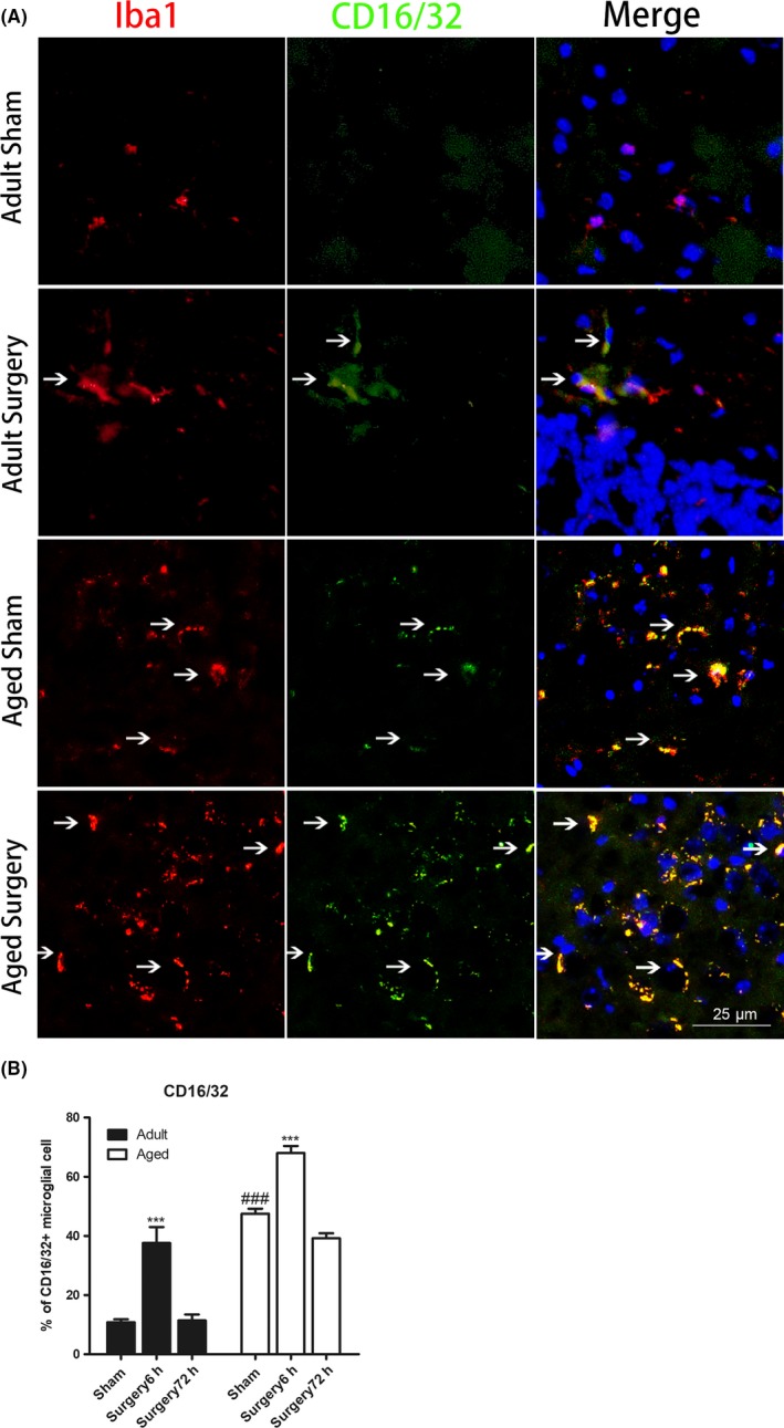
Figure 3.

Aging enhanced M1 microglial activation after peripheral surgery. A, Representative immunofluorescent images of the hippocampal dentate gyrus showing that aging enhanced M2 microglial activation at 6 h after peripheral surgery (scale bar, 25 μm). Brain sections were stained with Iba1 (red), M1 phenotype microglial marker CD16/32 (green), and DAPI (blue). B, Immunostaining analysis showing that the percentage of CD16/32‐positive microglia was higher in aged sham as compared with adult sham‐operated mice. Following tibial fracture, the percentage of CD16/32‐positive microglia significantly increased at 6 h and returned to baseline level 72 h after surgery in both adult and aged mice. All data are presented as the mean ± SEM. n = 4‐5; ***P < .001 vs age‐matched sham; ###P < .001, vs adult sham, two‐way ANOVA followed by Bonferroni's post hoc test
