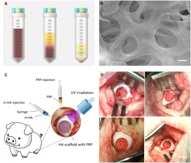
Figure 1.
(A) Schematic of the preparation of autologous PRP (left, collection of whole blood; middle, first centrifugation; right, second centrifugation). (B) The SEM of m-HA hydrogel. Scale bar: 5 µm. (C) Schematic of surgical procedure. (D) The demonstration of FT cartilage defect and osteochondral defect in the medial femoral condyle. Scale bar: 5 mm (top, FT cartilage defect; bottom, osteochondral defect)

