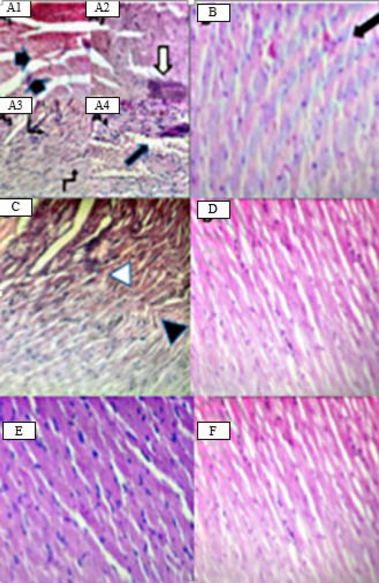
Fig. 5.

Representative hematoxylin and eosin (H&E) histological sections of the cardiac tissue of (A) DOX showing rupture of cardiac muscle fibers (black arrows in A1), hemorrhage (white arrow in A2) myocyte wavy degeneration (angled arrows in A3), necrosis and inter-fibrillar congestion (black arrow in A4). (B) Dexrazoxane (50 mg/kg) showing mild hemorrhage (black arrow). (C) Vanillic acid at dose of 10 mg/kg showing mild inter-fibrillar congestion (white arrowhead) and mild wavy degeneration (black arrowhead). (D and E) Vanillic acid at doses of 20 and 40 mg/kg and (F) normal saline control group, showing normal cardiac tissue architecture; ×40 magnification.
