Figure 5.
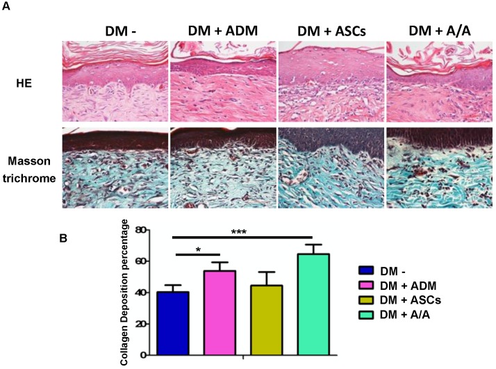
Collagen deposition significantly increased by ASCs-ADM. (A) Representative image of H&E and Masson trichrome staining, and (B) collagen deposition percentage of the group with the DM wound treated with ADM alone (DM + ADM) was significantly higher (p < 0.05) than that of the group with the diabetic wound without treatment (DM-). The group with the DM wound treated with ADM-ASCs (DM + A/A) showed a significantly higher (p < 0.001) collagen deposition percentage compared with the group with the DM wound without treatment (DM-). Scale bar = 50 μm.

