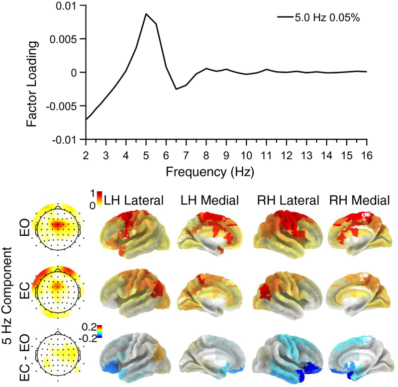
Figure 4.
Midfrontal theta component extracted by combined CSD/eLORETA-fPCA. A. Factor loadings revealed a peak frequency at 5 Hz. B. The overall component topography was consistent with previous work investigating midfrontal theta. Component tomography suggests sources in premotor areas, including the dACC. There were no significant condition differences (EC vs EO) for the theta component after multiple comparisons correction (corrected ps > .3). LH = Left Hemisphere; RH = Right Hemisphere.

