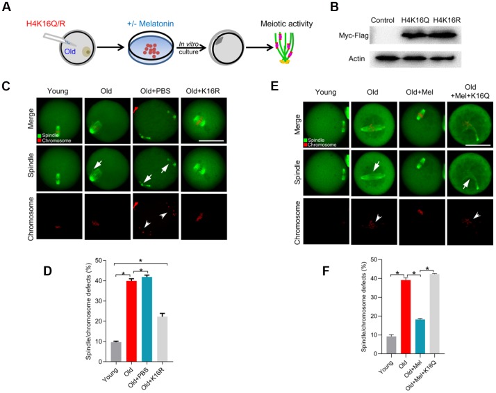
Figure 6.
Overexpression of H4K16R mutant ameliorates the maternal age-associated meiotic defects in mouse oocytes. (A) Schematic illustration of the experimental protocol to check whether H4K16 acetylation mediates the effects of melatonin on the quality of aged oocyte. (B) Western blotting showing that two mutant H4K16 proteins were expressed to the similar extent. (C) Young, old, old+PBS and old+H4K16R oocytes were stained with α-tubulin to visualize spindle (green) and counterstained with propidium iodide to visualize chromosomes (red). Representative confocal sections are shown. Arrowheads indicate the misaligned chromosomes and arrows indicate the abnormal spindles. (D) Quantification of young, old, old+PBS and old+H4K16R oocytes with spindle/chromosome defects. (E) Young, old, old+Mel and old+Mel+H4K16Q oocytes were stained with α-tubulin to visualize spindle (green) and counterstained with propidium iodide to visualize chromosomes (red). Representative confocal sections are shown. Arrowheads indicate the misaligned chromosomes and arrows indicate the abnormal spindles. (F) Quantification of young, old, old+Mel and old+Mel+H4K16Q oocytes with spindle/chromosome defects. Data are expressed as mean percentage ± SD from three independent experiments in which at least 100 oocytes were analyzed. *P<0.05 vs. controls. Scale bars: 50 μm.

