Figure 6.
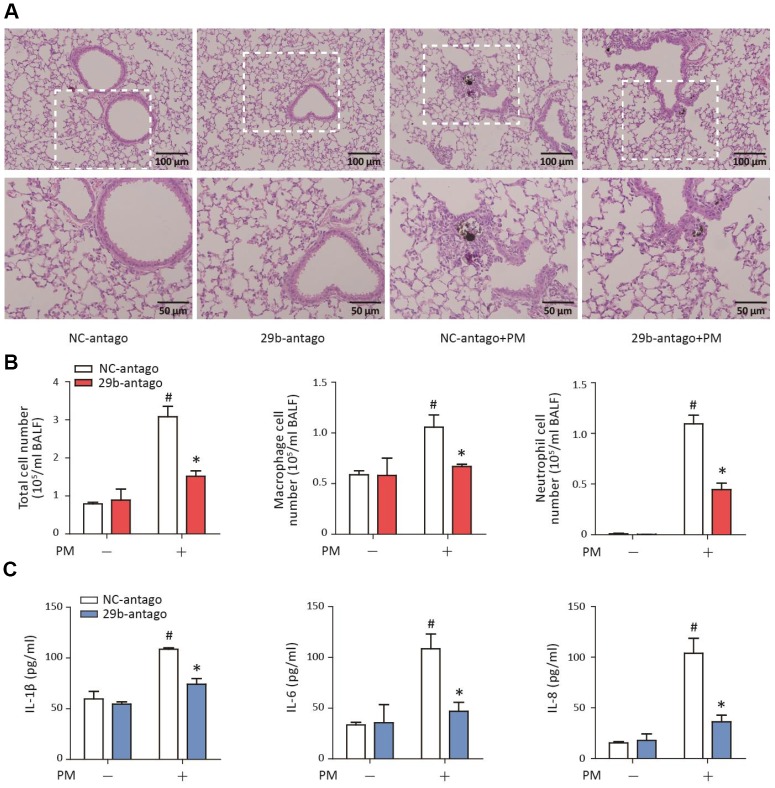
MiR-29b-3p inhibition attenuated the PM-induced acute lung inflammatory responses in vivo. The acute PM-exposed mouse model was constructed and miR-29b-3p antagomirs (29b-antago) or negative control antagomirs (NC-antago) were delivered via the tail vein of mice 24 h prior to the first PM exposure. (A) The histopathologic analysis of acute inflammatory responses in lung tissue of mice using H&E staining. (B) The numbers of total cells, macrophages and neutrophils in the BALF of mice were counted. (C) The concentrations of IL-1β, IL-6, and IL-8 in the BALF of mice were measured by ELISA. Values represent mean ± SEM; *, P<0.05, compared with the NC-antago + PM group; #, P<0.05, compared with the NC-antago group; n=3. BALF, bronchoalveolar lavage fluid; PM, particulate matter.

