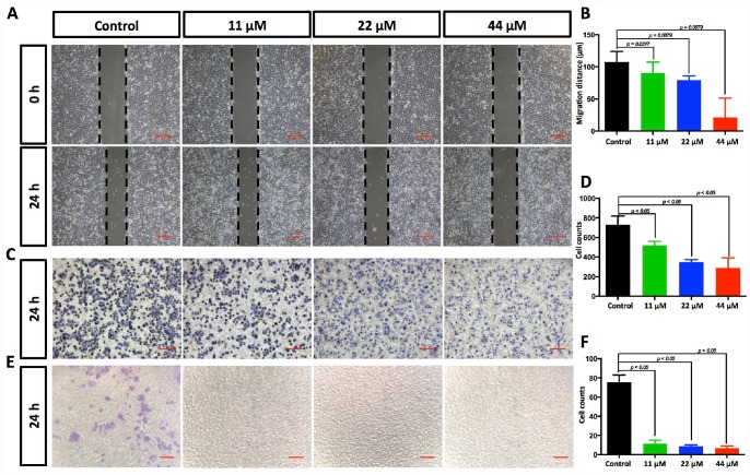
Figure 2.
TPGS dose dependently restrained HCC cell migration and invasion. (A) Effects of TPGS treatments on HCC cell migration, scale bar = 100 μm (B) The migration distance of HCC cells was quantified by ImageJ software, and the 44 μM TPGS group had the shortest migration distance (23 μm). (C) The inhibition of HCC cell migration by TPGS was confirmed by Transwell assays, scale bar = 100 μm. (D) The migrated cells were counted after Crystal violet staining with the 44 μM TPGS group having the lowest number of migrated cells (approximately 298). (E) TPGS diminished cell invasion of HCC cells (Transwell assay using an 8 μm pore filter coated with 0.5 mg/mL Matrigel), scale bar = 100 μm. (F) The mean cell counts of invading cells, with the 44 μM TPGS group having the lowest number of invasion cells (approximately 6).

