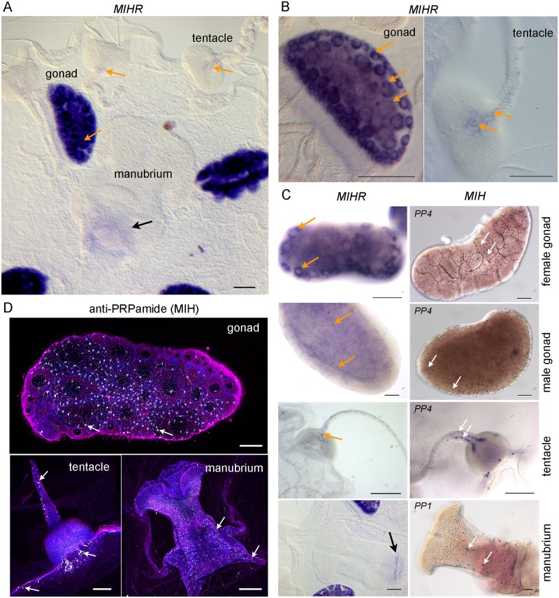
Fig 3. Sites of MIHR expression in the Clytia medusa.
(A) In situ hybridization detection of MIHR mRNA in a young adult Clytia female medusa. Strong purple MIHR signal (orange arrows) was detected in oocytes within the gonads, as well as in scattered cells in tentacles. (B) Higher magnification images of MIHR mRNA detected in young adult medusae; with the gonad, each oocyte (e.g., at orange arrows) has an unstained nucleus. The intensity of labelling in individual oocytes decreases as they grow due to dilution of cytosol with yolk. In the tentacle, a row of individual MIHR-positive putative neural cells can be clearly distinguished leading from the center of the bulb on its oral face. (C) Comparison of the distribution of the MIH receptor and ligand expressing cells in different medusa structures as labelled, detected by in situ hybridization using probes to MIHR (top row) and to the MIH peptide precursors PP1 and/or PP4 as indicated (bottom row). Orange arrows point to oocytes, developing spermatozoa and tentacle MIHR cells, and white arrows indicate MIH cells. The focal plane in the male gonad image is through the center to illustrate the position of the MIH cells in the ectodermal layer. Weak staining at the base of the manubrium (black arrow) in A and B is frequently observed with probes for many genes and is probably due to a specific trapping of the color reagent. Scale bars: 100 μm. (D) Confocal images of the three main sites of MIH-expressing cells (white arrows) in medusae, visualized using anti-PRPamide antibody (MIH: white), anti-tyrosinated tubulin (magenta), and Hoechst staining of nuclei (blue). Summed z-stacks are shown in all cases except for the gonad tubulin and DNA staining, where a single plane was selected through the center of the gonad. All scale bars: 100 μm. MIH, maturation-inducing hormone; MIHR, MIH receptor.

