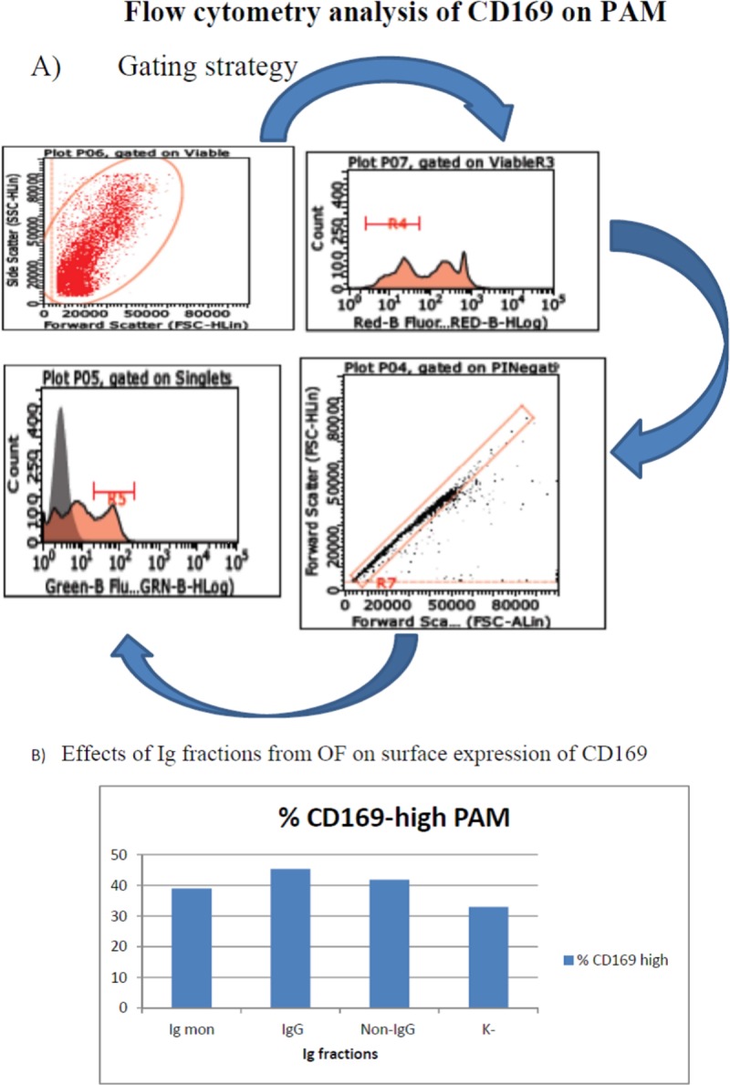
Fig 6. Surface expression of CD169 in PAM, test 4.

The direct interaction of IgG, monomeric IgA+IgG and non-IgG fractions with PAM was investigated. Panel A: gating strategy. PAM were gated first by a combination of forward and side scatter. Next, viable cells were selected by staining with PI (red fluorescence), and submitted to singlet analysis. CD169+ PAM were detected by monoclonal antibody 3B11/11 and Alexa Fluor® 488 F(ab')2 fragment of goat anti-mouse IgG, IgM (H+L). Panel B: prevalence of CD169-high PAM is depicted in a bar graph. The three Ig fractions caused up-regulation of CD169 in PAM (P< 0.001 with respect to control cells).
