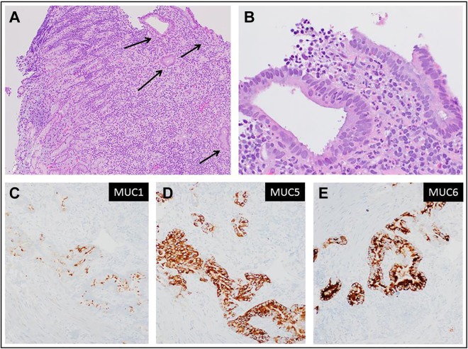
Figure 4.
Metastatic/recurrent pancreatic adenocarcinoma to gastric wall. A, Gastric biopsy; low magnification showing a fragment of gastric mucosa with unremarkable superficial gastric epithelium and underlying lamina propria. At the far right, malignant glands invade the lower part of the lamina propria (hematoxylin-eosin, original magnification ×40). B, Higher magnification showing medium-sized glands with haphazard growth, composed of malignant cells with marked variation in nuclear size, disorderly arrangement of nuclei, irregular nuclear membranes (hematoxylin-eosin, original magnification ×400). C-E, Immunohistochemical stain for mucin-1, mucin 5, and mucin 6, respectively, highlighting the malignant glands (original magnification ×200).

