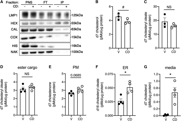
Fig. 2.
Lysosomal cholesterol is differentially distributed to the media and the ER after CD treatment. U2OS-SRAshNPC1 cells that express epitope-tagged lysosomal protein TMEM192 (A–C) or U2OS-SRAshNPC1 cells (D–G) were incubated overnight with d7-acLDL in the presence of lalistat and then treated for 6 h in media with CD or vehicle before the immunoisolation of intact lysosomes (A–C) or analysis of other compartments (D–G). A: Immunoblots of PNS, FT, and IPs for LMP, HEX, CAL, COX, HIS, and NAK. B, C: LC/MS/MS quantification of lysosomal d7 cholesterol (B) and d7 cholesteryl oleate (C) normalized to lysosomal protein. D–G: d7 cholesterol species quantified by LC/MS/MS in treated cells that also received d9 oleate. Remaining d7 cholesteryl ester cargo (D), PM d7 cholestenone (E), ER reesterification product d16 cholesteryl oleate (F), and media-associated d7-cholesterol (G), each normalized to cellular protein. Means ± SEs (n = 3–4). #P < 0.05 by paired t-test (B, C); *P < 0.05 by unpaired t-test (D–G). CAL, calreticulin; COX, cytochrome c oxidase IV; FT, flow through; HEX, hexaminidase; HIS, histone 3; IP, immunoisolated lysosome; LMP, LAMP1; NAK, NaK ATPase; PNS, postnuclear supernatant.

