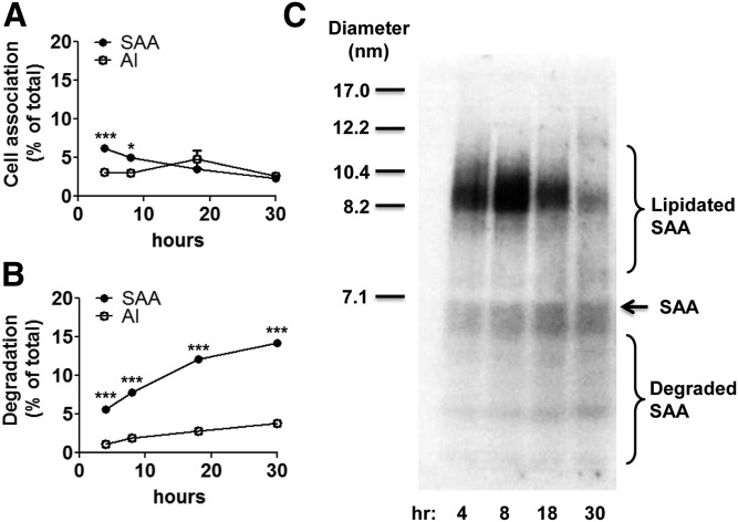
Fig. 2.
Exogenous SAA is degraded by hepatocytes to a greater extent than apoA-I. Primary hepatocytes were prepared from SAA1.1/2.1/3-TKO mice as described in the Materials and Methods and incubated at 37°C for the indicated times with 5 μg/ml 125I-SAA or 125I-apoA-I. A: After removing the culture media, cells were solubilized in 0.1 N NaOH, and the protein and 125I contents of the lysates were determined and expressed as the percentage of the total amount of 125I added to the dish. B: The cell-mediated degradation of SAA and apoA-I was measured in the culture media and expressed as the percentage of the total amount of 125I added to the dish. Statistical analysis was performed using two-way ANOVA with Bonferroni’s multiple comparison tests. P-values less than 0.05 and 0.001 are denoted with one (*) and three asterisks (***), respectively. C: Aliquots of the cell media at each time point were separated by NDGGE followed by autoradiography to visualize lipidated and degraded SAA. The migration of size standards, lipidated SAA, lipid-free SAA, and SAA degradation products are indicated.

