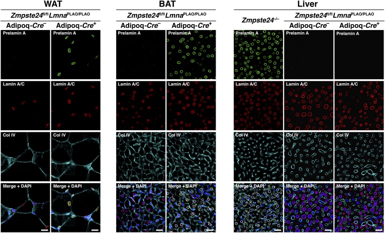
Fig. 2.
Prelamin A accumulates in adipocytes of LmnaPLAO/PLAOZmpste24fl/flAdipoq-Cre+ mice, as judged by immunofluorescence microscopy. Tissue sections of gonadal WAT, BAT, and liver from a LmnaPLAO/PLAOZmpste24fl/flAdipoq-Cre– mouse and a LmnaPLAO/PLAOZmpste24fl/flAdipoq-Cre+ mouse were stained with antibodies against prelamin A (green), lamin A/C (red), and collagen IV (cyan). DNA was stained with DAPI (blue). Liver from a Zmpste24−/− mouse was included as a positive control for the prelamin A antibody. Images were recorded with a confocal microscope using a 20× objective and a 2× digital zoom. Identical microscope settings were used for all three tissue samples. Scale bar, 20 μm.

