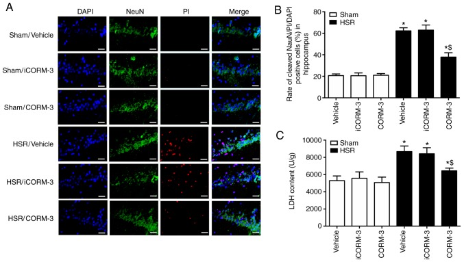
Figure 4.
CORM-3 ameliorates HSR-induced neuronal death in the hippocampus. (A) Representative photomicrographs of NeuN/PI/DAPI staining (NeuN, green; PI, red; DAPI, blue) demonstrating neuronal death in the hippocampal tissue induced by the indicated stimuli. Scale bar, 50 µm. (B) Percentages of neuronal death in the hippocampal tissue induced by the indicated stimuli. (C) LDH content in the hippocampal tissue induced by the indicated stimuli. Data are presented as the mean ± SD (n=6 per group). *P<0.05 vs. Sham; $P<0.05 vs. HSR vehicle and HSR iCORM-3. CORM, carbon monoxide-releasing molecule; HSR, hemorrhage shock and resuscitation; iCORM-3, inactive CORM-3; PI, propidium iodide; LDH, lactate dehydrogenase.

