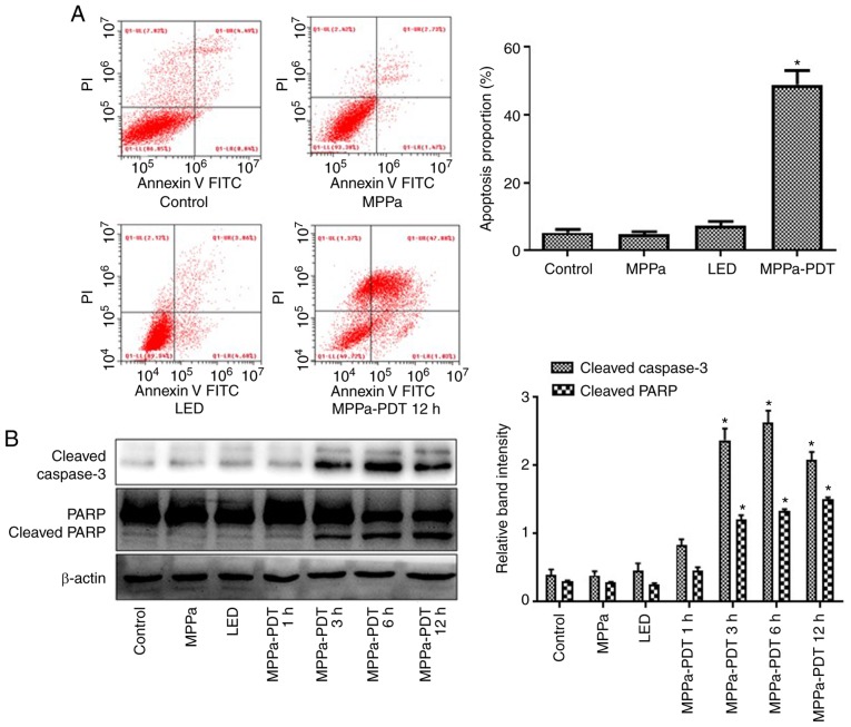
Figure 4.
MPPa-PDT induces MG-63 cell apoptosis. (A) Following MPPa-PDT treatment for 12 h, apoptotic rates were analyzed by flow cytometry and calculated as the percentage of early apoptotic (Annexin V+/PI−) cells plus the percentage of late apoptotic (Annexin V+/PI+) cells. (B) At 12 h, whole-cell lysates were prepared to determine cleaved caspase-3 and cleaved PARP protein expression by western blotting. Data are presented as the mean ± SD from three independent experiments. *P<0.05 vs. control. MPPa, pyropheophorbide-α methyl ester; PDT, photodynamic therapy; LED, cells treated with a light-emitting diode; PI, propidium iodide; PARP, poly (ADP-ribose) polymerase 1.

