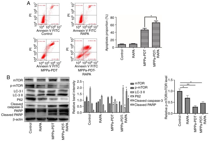
Figure 6.
Apoptosis is promoted via mTOR to enhance MPPa-PDT-induced autophagy in MG-63 cells. MG-63 cells were pretreated with RAPA for 3 h in the presence or absence of MPPa-PDT. Control cells received no pretreatment or only RAPA pretreatment. (A) Following the corresponding treatment, apoptotic rates were measured by flow cytometry. The apoptotic rate was calculated as the percentage of early apoptotic (Annexin V+/PI−) cells plus the percentage of late apoptotic (Annexin V+/PI+) cells. (B) At 12 h after treatment, whole-cell lysates were prepared to determine p-mTOR, LC-3, P62, cleaved caspase-3 and cleaved PARP expression by western blotting. Data are presented as the mean ± SD from three independent experiments. *P<0.05 and **P<0.01. vs. MPPa-PDT. MPPa, pyropheophorbide-α methyl ester; PDT, photodynamic therapy; RAPA, rapamycin; PI, propidium iodide; PARP, poly (ADP-ribose) polymerase 1; LC-3, microtubule-associated protein 1 light chain 3α; p, phosphorylated.

