Figure 3.
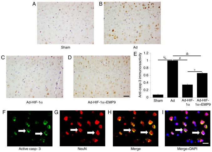
Ad-HIF-1α treatment suppressed active caspase-3 expression in neurons. Active caspase-3 was detected in (A) the Sham group, (B) the Ad group, (C) the Ad-HIF-1α and (D) the Ad-HIF-1α +EMP9 group (scale bar, 50 µm), and then (E) quantified. There were significantly more active caspase 3-positive cells in the brains of rats treated with Ad compared with the Sham group. %P<0.05, #P<0.05, &P<0.05 and *P<0.05 with comparisons shown by lines. (F) Active caspase 3 and (G) NeuN double-labeled fluorescence staining in Ad-treated rat brains (scale bar, 20 µm). (H) Colocalization of NeuN (red) and active caspase 3 (green) (white arrows; scale bar, 20 µm). (I) Colocalization of NeuN (red), and active casp3 (green) and DAPI (blue) (white arrows; scale bar, 20 µm). Ad, adenovirus; HIF-1α, hypoxia-inducible factor-1α; EMP9, erythropoietin mimetic peptide-9; NeuN, RNA binding fox-1 homolog 3.

