. 2020 Feb 20;9:e53515. doi: 10.7554/eLife.53515

Figure 3. A closed DNA substrate is required for an increased association of ParB with DNA.

(A) BLI analysis of the interaction between a premix of 1 μM Caulobacter ParB-His6 ± 1 mM CTP and a 169 bp dual biotin-labeled parS DNA. (B) Same as panel A but immobilized DNA fragments have been restricted with BamHI before BLI analysis. (C) Same as panel A but immobilized DNA fragments have been restricted with EcoRI before BLI analysis. Schematic of DNA fragments with the relative positions of parS and restriction enzyme recognition sites are shown above the sensorgram. Each BLI experiment was triplicated and a representative sensorgram was presented. (D) A schematic of the pull-down assay and immunoblot analysis of pulled-down Caulobacter ParB-His6. For lanes 3 and 4, DNA-bound beads were incubated with HindIII to linearize bound DNA. Samples in lanes 1–4 were loaded on the same gel, the immunoblot was spliced together for presentation purposes. All buffers used for experiments in this figure contained Mg2+.

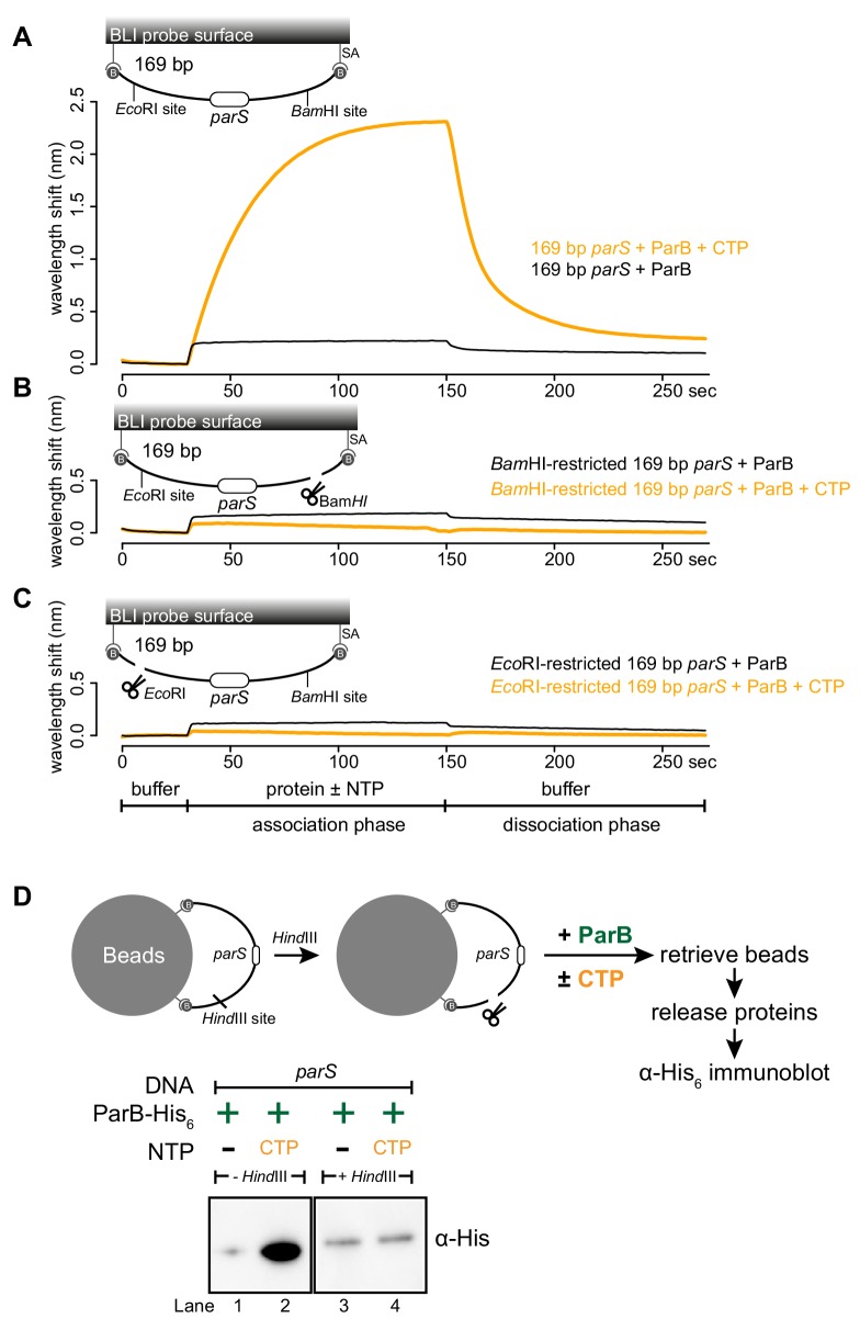
Figure 3—source data 1. Data used to generate Figure 3.

Figure 3.

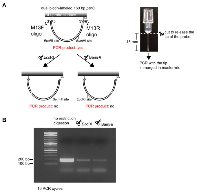
Figure 3—figure supplement 1. Restriction enzymes linearize dual biotin-labeled DNA fragments on the surface of the BLI probe.

Figure 3—figure supplement 1.

(A) (Left panel) A schematic of a digestion assay using BamHI/EcoRI and PCR. PCR was performed using M13F, M13R oligos, and DNA attached to the BLI surface as a template. (Right panel) The BLI probe was severed from the plastic adaptor and immerged into a PCR master mix. (B) Detection of intact 169 bp DNA fragments that were bound on the surface of the BLI probe before and after treatment with restriction enzymes.
Figure 3—figure supplement 1—source data 1. Data used to generate Figure 3—figure supplement 1.
Figure 3—figure supplement 2. Linearization of a closed DNA substrate by BamHI liberates pre-bound ParB from DNA.

Figure 3—figure supplement 2.

BamHI was added after 1 μM Caulobacter ParB-His6 + 1 mM CTP and a 169 bp dual biotin-labeled parS DNA were preincubated together for 120 s. As a negative control, heat-inactivated BamHI was added instead. Mg2+ was included in all buffers for this experiment. A schematic of the DNA substrate is shown above the sensorgram.
Figure 3—figure supplement 2—source data 1. Data used to generate Figure 3—figure supplement 2.