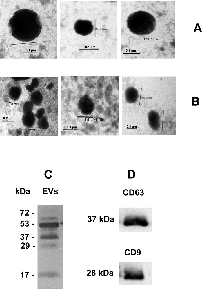
Fig 3. Images captured by transmission electron microscopy.
Negatively stained microvesicles and exosomes of cerebrospinal fluid (Panel A) and serum (Panel B) purified by ultracentrifugation. Barr = 100 nm and 200 nm. Magnification of all images, 150, 000. Serum-derived EVs were separated by 10% SDS-PAGE and stained by silver. (Panel C). Next, serum-derived EVs, separated by 10% SDS-PAGE was transferred to nitrocellulose and incubated with anti-CD63 and anti-CD9 antibodies (Panel D).

