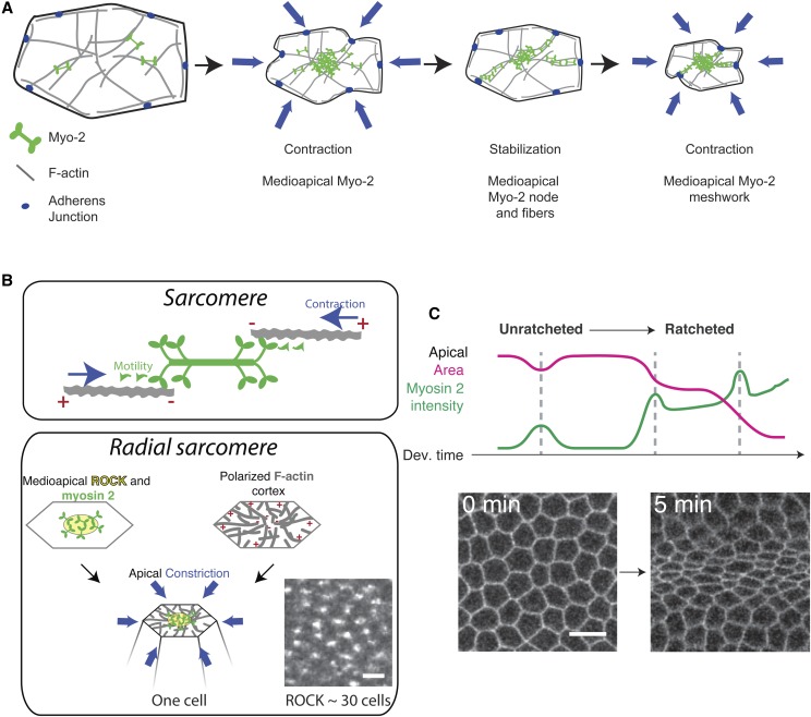
Figure 4.
Spatial and temporal organization of myosin 2 contractility in mesoderm cells. (A) Ratcheted apical constriction of mesoderm cells. Medioapical actomyosin pulls centripetally on adherens junctions in stepwise constriction of the cell apex. (B) Apical cortex has a radial organization. Top, myosin 2 interacting with antiparallel actin filaments with plus ends facing out enables filament sliding and contraction. Bottom, medioapical myosin 2 activation and radial organization of actin filaments with outward facing plus ends enable actin network to be pulled toward center, constricting the apex. Image shows ROCK localization during apical constriction of the mesoderm, which shows a clear periodic pattern across the tissue—each spot represents the center of a contractile unit, which is a single cell. Bar, 3 μm. (C) Temporal progression of myosin 2 pulsing. Initially unratcheted pulses occur and then cells transition to having ratcheted pulses, which leads to sustained changes in apical area. Images show unconstricted and then constricted cell apices in the process of invagination. Images are reproduced from Chanet et al. (2017). Bar, 10 μm.

