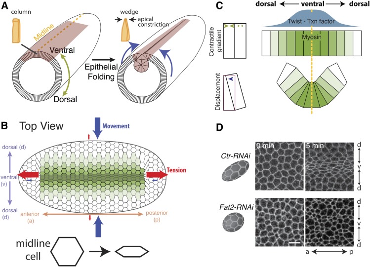
Figure 6.
The mechanics of Drosophila mesoderm invagination. (A) Cartoon showing cell and tissue shape changes during mesoderm invagination. Orange cells illustrate cell shape. Blue arrows show movement of ectoderm tissue during mesoderm invagination (maroon). Note that ventral side is shown facing up to highlight the mesoderm. (B) Birds-eye view of presumptive mesoderm. Green illustrates the level of cell contractility. Red arrows denote tension and blue arrows denote movement. (C) Cross-section view of mesoderm cells during invagination. A gradient in twist expression (blue curve) results in a gradient in myosin 2 contractility (green), which promotes efficient apical constriction in the ventral–dorsal direction. (D) Apical constriction anisotropy depends on embryo shape. Images are subapical views of cells showing cell outlines. In the control, ellipsoidal embryos (Ctr-RNAi), cells constrict mostly in the ventral–dorsal direction. In round embryos (Fat2-RNAi), cell constrict isotropically. Images are reproduced from Chanet et al. (2017). Bar, 10 μm.

