Figure 3.
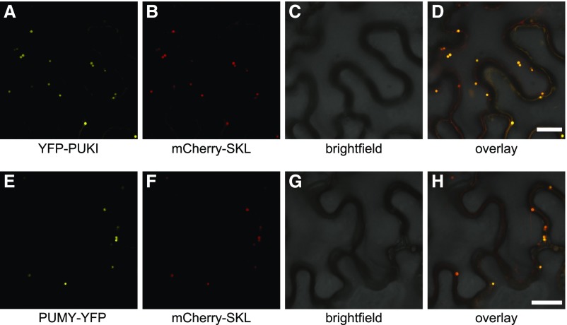
Subcellular Localization of PUKI and PUMY.
(A) to (D) Confocal fluorescence microscopy images of N. benthamiana cells in the lower leaf epidermis transiently coproducing mCherry-SKL and PUKI fused to an N-terminal YFP tag (YFP-PUKI): YFP (A), mCherry (B), brightfield (C), and overlay of YFP, mCherry, and brightfield (D). All infiltrated cells exhibited the same pattern. Bar = 25 µm.
(E) to (H) Same as in (A) to (D) but for C-terminally YFP-tagged PUMY.

