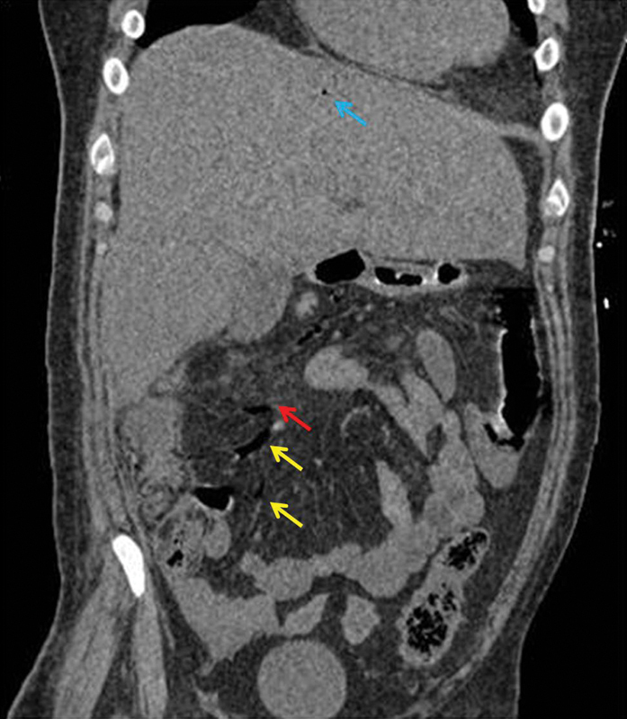
Fig. 3.

Coronal noncontrast computed tomography (CT) scan image showing air in the ileocolic vein ( yellow arrow ) and the right colic vein ( red arrow ), and hepatic portal venous gas ( blue arrow ). Also, air in the small tributaries of the superior mesenteric vein can be seen.
