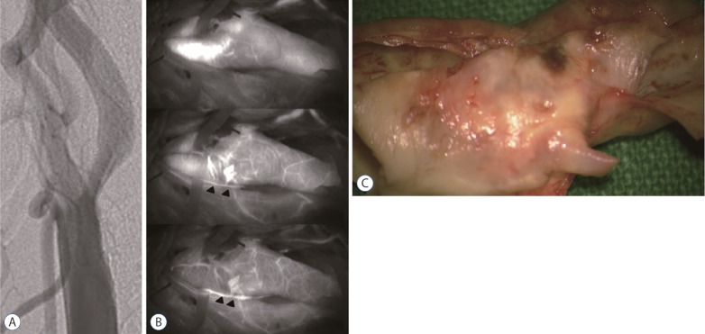
Fig. 4.
Representative case of vasa vasorum venosum (VVV). A 64-year-man presented with a recurrent a transient ischemic attack on the left side with a history of smoking. A : Carotid artery angiogram showing carotid stenosis (60%, the North American Symptomatic Carotid Endarterectomy Trial). B : Indocyanine green-video angiography image demonstrating delayed depiction of the VVV (arrowheads) along with the carotid bulb. C : Intraoperative findings indicating stable plaque—smooth outer and inner surfaces.

