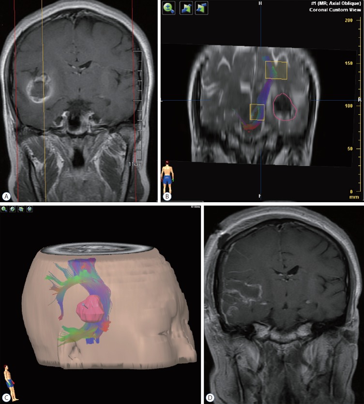
Fig. 2.
Illustrative case 1. A 29-year-old woman presented to our service, having experienced a seizure and subsequent severe headache. A : Preoperative magnetic resonance imaging revealed a cavernous malformation of right temporal lobe, with pial presentation. B : Diffusion tensor imaging fiber tracking indicated an intact corticospinal tract, albeit distorted along the central aspect of lesion; and there was a 10-mm distance from presumptive pial border to corticospinal tract. The red circle delineated the tumor border. C : The relation between tumor and corticospinal tract was defined, using our neuronavigation system for surgical trajectory planning. D : Postoperative magnetic resonance imaging showed complete eradication of the lesion.

