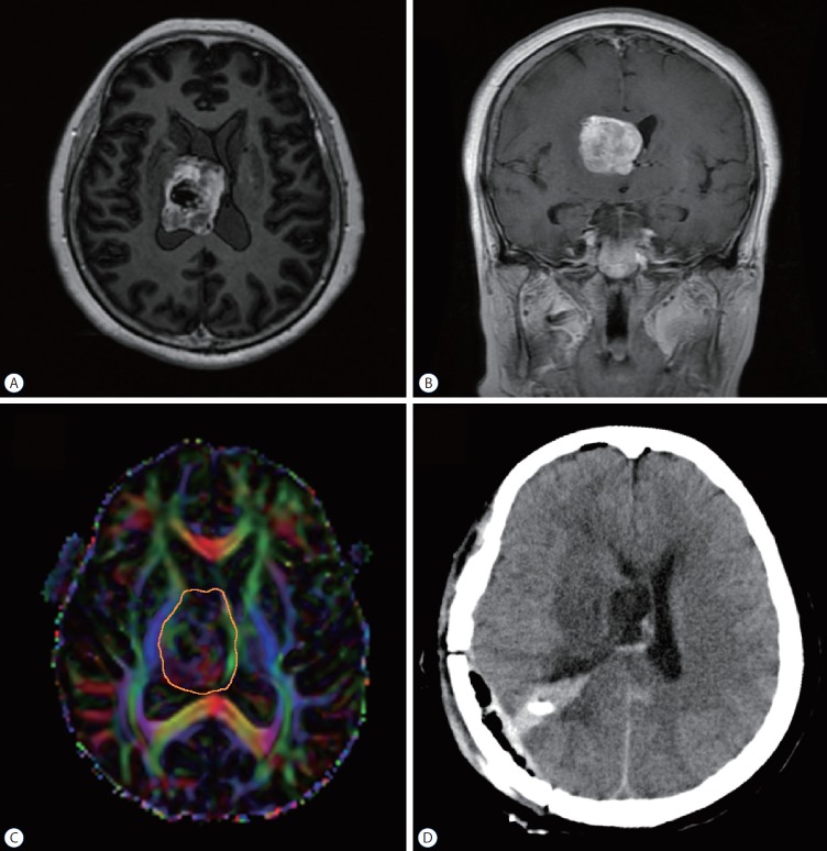
Fig. 3.
Illustrative case 2. The patient was a 67-year-old woman who complained about left leg weakness accompanied with blurred vision in both eyes for 2 months. A and B : Preoperative magnetic resonance imaging revealed a large mass situated in the right ventricle. C : DTI analysis showed that the tumor was closely located inside the CST. The tumor-CST was 0 mm (C). Comparing to the CST in the counterpart hemisphere, the integrity of CST bundle was intact but distorted laterally (simple displacement). The orange circle delineated the tumor border. D : After DTI and neuronavigation analysis, we performed a tumor resection through transinferior parietal approach. The tumor was completely removed under the neuronavigation guidance. DTI : diffusion tensor imaging, CST : corticospinal tract.

