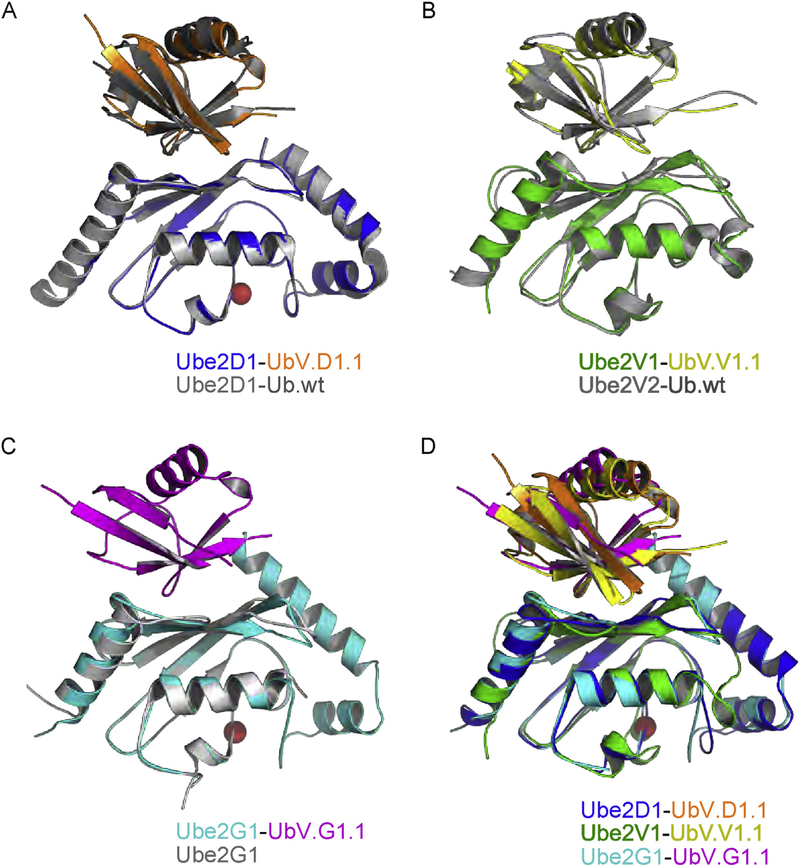
Fig. 3. Superpositions of structures of UbVs and Ub.wt in complex with E2 proteins.
Ub.wt and its associated E2 protein are colored gray. The UbV and its associated E2 protein are colored as follows: UbV.D1.1 (orange) and Ube2D1 (blue), UbV.V1.1 (yellow) and Ube2V1 (green), UbV.G1.1 (magenta) and Ube2G1 (cyan). Active site cysteines in E2 enzymes are shown as red spheres. (A) Superposition of Ube2D1-UbV.D1.1 complex with Ube2D1-Ub.wt complex (PDB: 3PTF). (B) Superposition Ube2V1-UbV.V1.1 complex with Ube2V2-Ub.wt complex (PDB: 1ZGU). (C) Superposition of Ube2G1-UbV.G1.1 complex with apo Ube2G1 structure (PDB: 2AWF). (D) Superposition of Ube2D1-UbV.D1.1, Ube2V1-UbV.V1.1 and Ube2G1-UbV.G1.1 complexes.

