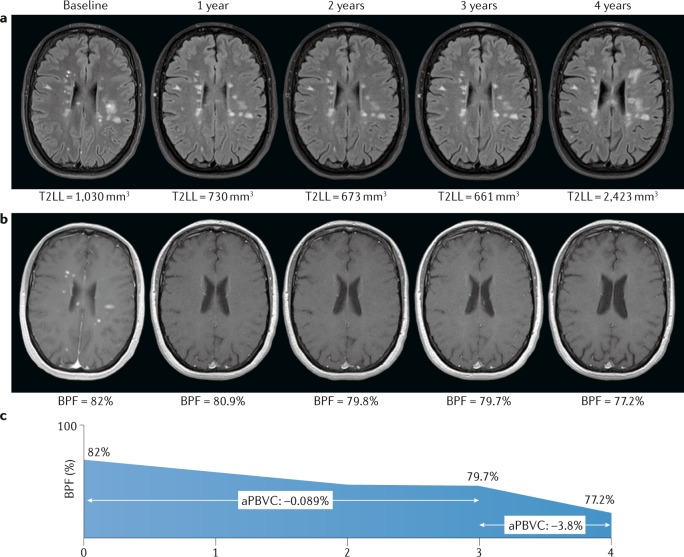
Fig. 1. Lesion load and brain atrophy in relapsing–remitting multiple sclerosis.
a | Transverse T2-weighted fluid attenuation inversion recovery images from a patient with highly active relapsing–remitting multiple sclerosis (MS) who started a disease-modifying therapy at baseline. The T2 lesion load (T2LL) is stable during the first 3 years of treatment while the patient remained clinically stable (no relapses and no disability worsening), but markedly increases at the fourth year after treatment discontinuation associated with clinical activity (the rebound effect). b | Contrast-enhanced T1-weighted images from the same patient showing the change in brain parenchymal fraction (BPF) over time. The decrease in global brain volume in the first 3 years is mild (annualized percentage of brain volume change (aPBVC) −0.089%), but the volume loss at the fourth year is severe (aPBVC −3.8%), matching the change in T2LL and clinical evolution. The severe loss observed in year 4 is well beyond the −0.4% suggested as a pathological cut-off for brain volume loss in MS24. c | Graphical representation of the changes in BPF over time, emphasizing the dramatic loss of volume in year 4.

