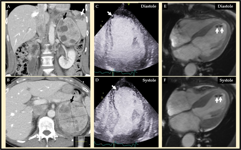
Fig. 1.
Abdominal computed tomography (CT) (a and b) revealed a left adrenal tumor, which turned out to be a pheochromocytoma (black arrow), and a rupture of spleen (A white arrow) with retroperitoneal bleeding. Contrast echocardiography (c diastole and d systole) revealed mid-apical ballooning typical for takotsubo syndrome with an apical filling defect suggestive of left ventricular thrombus (white arrow). Cardiac magnetic resonance (CMR) imaging (e diastole and f systole) confirmed the mid-apical pattern of takotsubo syndrome and the apical left ventricular thrombus (white arrows)

