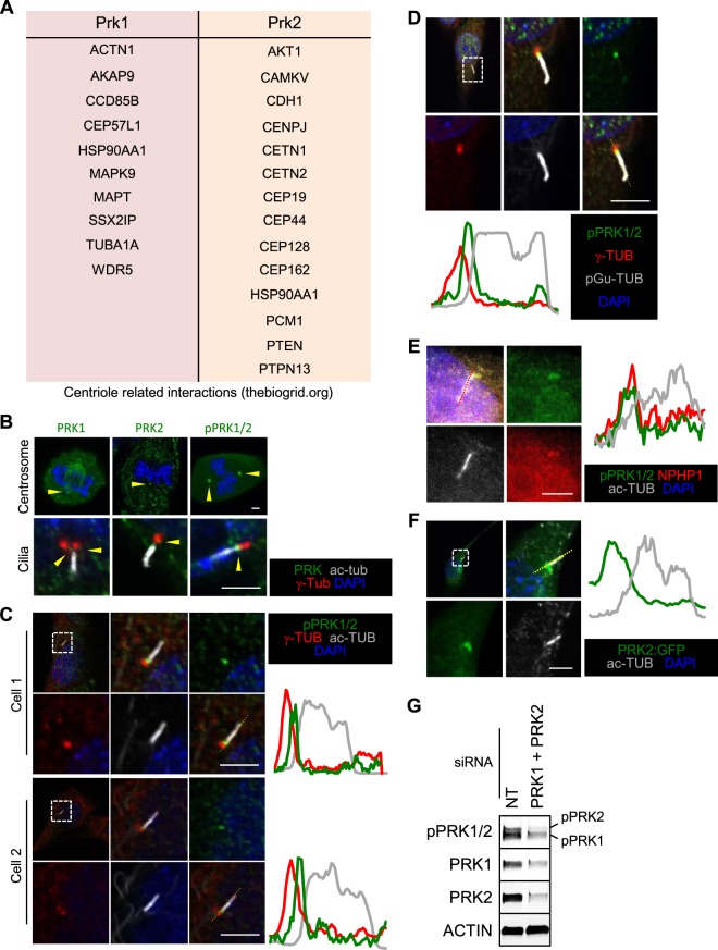
Figure 1.
PRK1 and PRK2 interact with centriole associated components and structures and localise to the transition zone of cilia. (A) Centriole associated proteins stated (thebiogrid.org) to interact with PRK1 or PRK2. (B) Immunofluorescence images of centriolar structures (centrosomes and cilia). (C–E) Immunofluorescence images of pPRK1 and pPRK2 (pPRK1/2) at the base of cilia with various other cilia associated markers. Line traces along the cilium are shown to demonstrate localization to the transition zone. (F) Immunofluorescence image of a NIH3T3 cell exogenously expressing PRK2 fused to GFP (PRK2:GFP). Scale bar represents 2 µm. (G) Western blot analysis of lysate from cells treated with non-targeting (NT) or PRK1 and PRK2 (PRK1 + PRK2) siRNA. Unless stated otherwise, n ≥ 3.

