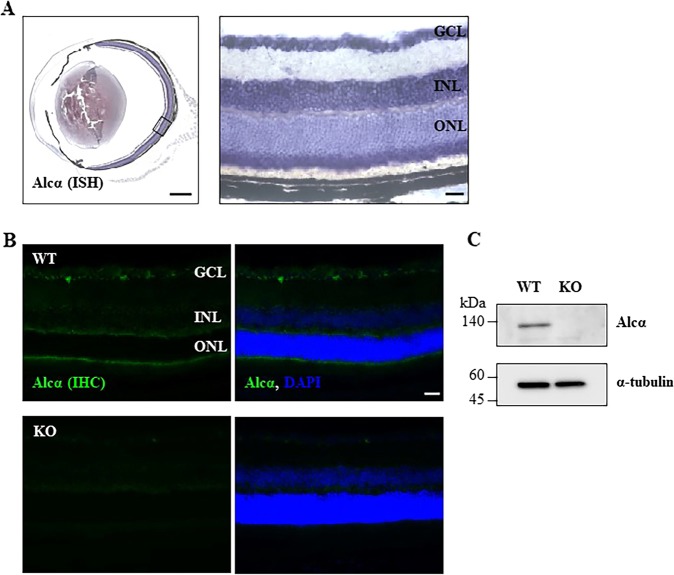
Fig. 1. Expression of Alcα in the mouse retina.
a Expression of Alcα mRNA in an eye of 5-month-old wild-type mice was detected by in situ hybridization (ISH). The magnified image in the boxed area of the left side image is shown on the right side. Alcα mRNA is readily detected in ganglion cells, cells in inner nuclear layer, and photoreceptors’ inner segment. GCL: ganglion cell layer, INL: inner nuclear layer, ONL: outer nuclear layer. Scale bar, 500 μm. b Expression of Alcα protein in an eye of 5-month-old wild-type (WT) and Alcα-deficient (KO) mouse was detected by immunohistochemistry (IHC). Alcα protein is readily detected in ganglion cells, photoreceptors’ inner segment, and outer peripheral layer. Alcα protein is not detected in the eye of Alcα-deficient mouse. Co-stained images with DAPI are shown on the right sides. GCL: ganglion cell layer, INL: inner nuclear layer, ONL: outer nuclear layer. Scale bar, 50 μm. c Expression of Alcα protein in a retina of 2-month-old wild-type (WT) and Alcα-deficient (KO) mouse was detected by western blotting. Alcα protein is not detected in the retinal lysate (1 μg) of Alcα-deficient (KO) mouse.

