Figure 8.
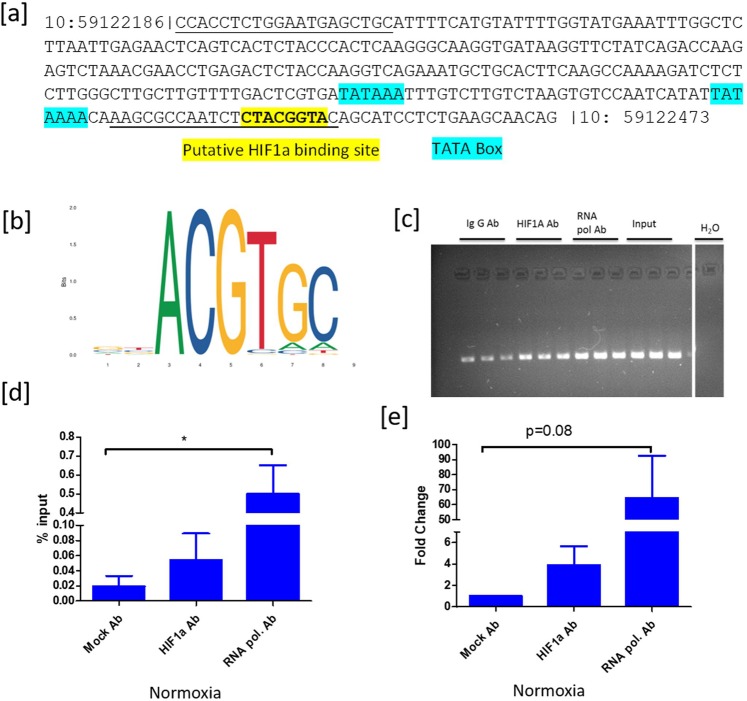
Chromatin immunoprecipitation analysis. (a) shows the nucleotide sequence between the physical location of 59122186 to 59122473 nucleotides on chromosome 10 in the bovine genome. Manually curated TATA box and the putative HIF1 binding sites were highlighted in blue and yellow colors. The primer binding sites were indicated with an underline. (b) adopted JASPAR Sequence logo of the HIF1 binding site based on ChIP seq data in humans. (c) indicates the agarose gel pictures of immune-precipitated and input DNA samples. (d,e) depict the relative quantification of HIF1 binding events on the CYP19A1 promoter based on percent input and fold change methods, respectively under FSH and IGF1 Supplemented conditions. Data were represented in mean ± SEM values of three independent experiments (n = 3). Significant differences were acknowledged at the minimum level of p < 0.05 by one way repeated measures analysis of variance. Pairwise comparisons were analyzed using Post hoc Tuckey test.

