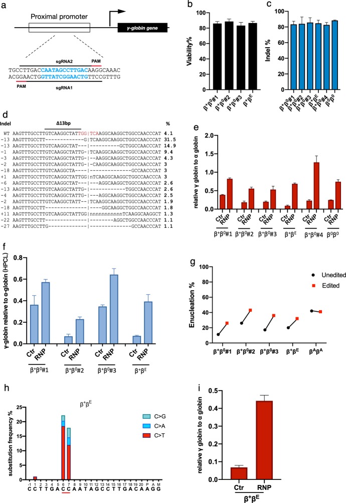
Fig. 1. Genome editing of HBG1/2 promoter reactivates the expression of γ-globin and ameliorates β-thalassemia.
a Schematic view of the targeted region in the HBG1/2 promoter. The 13-bp deletion in HPFH patient is labeled in blue. Spacers and protospacer adjacent motif (PAM) sequences are indicated with black and red lines, respectively. Black arrow indicates the direction of transcription. b Viability of the patient derived HSPCs 48 h after electroporation of the RNP complex. c Indel frequencies measured by TIDE analysis after RNP electroporation (data presented as mean ± SD, n = 3). d The most frequent indel patterns identified by TIDE analysis after sequencing the targeted site from the patient genomes. Frequency of each pattern is listed to the right. Red letters indicate the BCL11A-binding site. Δ13bp, 13-bp deletion site naturally occurred in HPFH patients. Horizontal dash lines indicate deletions and ‘n’ indicates substitutions. Vertical dash lines indicate the approximate cleavage site of Cas9. γ-globin level normalized to α-globin as measured by qPCR (e) or reverse HPLC (f) in patient HSPCs edited with Cas9/sg-HBG1 or Cas9 only (data presented as mean ± SD, n = 3). g The enucleation rate of edited or unedited patient HSPCs (data plotted as mean ± SD, n = 3). h C to D (A, T, or G) substitution frequencies induced by hA3A-BE3 in the targeted region in patient HSPCs. The −115C and −114C of the HBG promoter is underlined. i γ-globin level normalized to α-globin (qPCR) after targeting the HBG promoter by hA3A-BE3. β0β0 β-thalassemia major. β0β+ β-thalassemia intermedia. β+ βE β-thalassemia intermedia containing heterozygous E26K point on HBB gene. βA βA normal alleles.

